Description
Molecular Breast Imaging
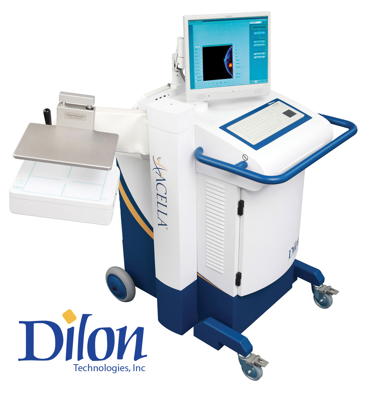
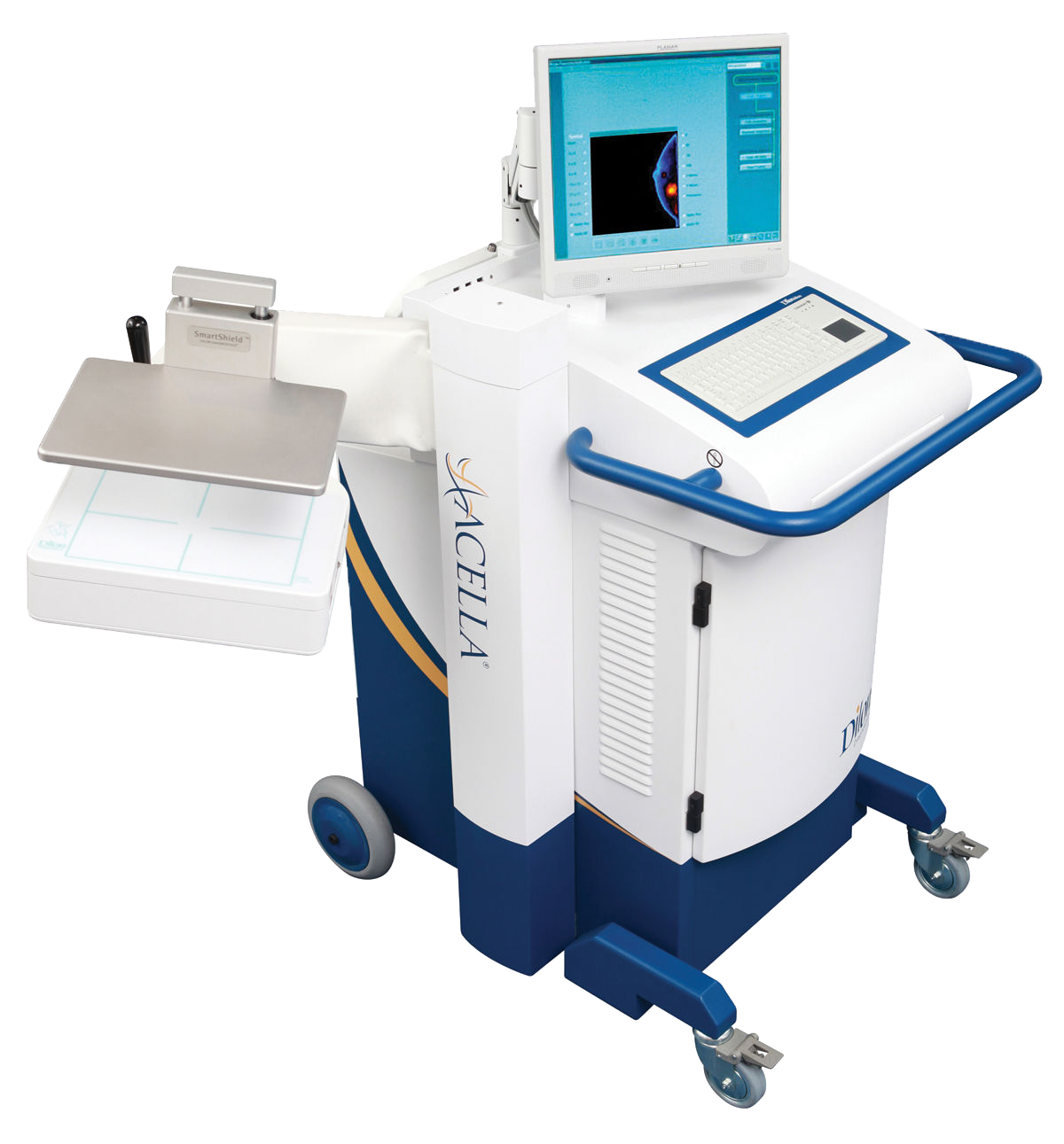
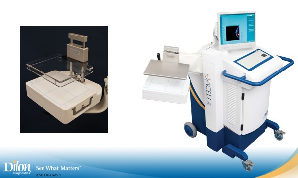
The Dilon Molecular Imaging System is a specialized camera designed to image various organs. This system has been optimized to perform Molecular Breast Imaging (MBI) and is a strong diagnostic tool in early breast cancer detection. Using MBI as an adjunct to mammography results in an almost fourfold increase in invasive cancer detection in women with dense breast tissue. MBI has a higher specificity than MRI and has proven to reduce benign biopsies by 50%. With a negative predictive value of 98% MBI is the beacon in dense breast tissue. The system offers MBI biopsy capability which provides an additional tool to accurately localize the region of interest.
The Dilon MBI procedure is an excellent problem-solving tool when faced with difficult-to-diagnose patients with:
• Dense Breasts
• Inconclusive Mammograms
• Lumps not seen by Mammogram
• Family History of Breast Cancer
• Breast Implants
• Breast Cancer Survivor
• Scar Tissue
• Cannot have an MRI
WHAT CAN MBI DO?
For the breast radiologist:
• Find cancers missed by mammography and ultrasound, especially in difficult and dense breast cases, while leading to fewer benign biopsies than MRI
• Provide a high negative predictive value to eliminate questionable findings on mammography and ultrasound
For the breast surgeon:
• Improve surgical planning by finding additional disease
• Provide a better understanding of the position of the tumor to help in surgical planning
• Provide information useful in monitoring tumor response to chemotherapy
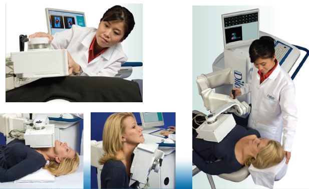
HOW DOES MBI WORK?
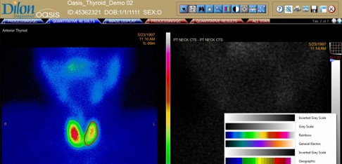
MBI uses a pharmaceutical tracer that is absorbed by the breast tissue. The cancerous cells in the breast absorb a greater amount of the tracer than normal, healthy cells. As a result, the tracer “lights up” the cancerous areas inside the breast and the malignant cells appear as “dark spots” on the MBI image. Patients find the exam to be very comfortable, unlike mammography, as there is minimal compression and they are seated throughout the process.
Its similarity to mammographic positioning makes the MBI an easy modality for any trained mammographer to learn and use, and allows easy comparison to the original mammography views by the radiologist. The breast cancer scanning procedure may begin five minutes after the patient has been injected with a small amount of radioactive tracer (Tc-99m Sestamibi). The entire breast cancer screening study takes approximately 40-45 minutes and images are immediately available for a physician’s interpretation.
MBI CLINICAL ASPECTS
The Dilon Molecular Breast Imaging (MBI) Procedure can detect cancers missed by mammography and ultrasound, while leading to fewer benign biopsies than MRI. According to clinical studies, MBI has very high sensitivity for identifying earlier stage cancers as small as 1 mm. Clinical indications of MBI include:
• Diagnosing breast cancer patients with an unresolved diagnostic dilemma
• Equivocal exams
• Dense-breast with indeterminate breast abnormalities
• Breast implants
• Discordant findings
• As an alternate to MRI in patients who can not have an MRI study
• Monitor for recurrence
• Evaluate the extent of disease (initial staging)
• Detect multi-centric, multi-focal, or bilateral disease
• Assess response to neo-adjuvant chemotherapy
• As an adjunct to a limited mammogram
• Surgical planning for residual disease
Dilon 6800: Technical Specifications
Digital Detector
Processing Mode
Design
Crystal Material
Overall Size
FOV and UFOV
Pixel Size
Crystal Thickness
Number of Crystals
Photomultiplier Tubes
Shielding
Dead space
Axes of Rotation
Detector Rotation
Detector Tilt
PSPMT & Pixilated Crystals
NaI[Tl]
10.9 x 25.1 x 29.2 cm
[4.3 x 9.9 x 11.5 in]
15.2 x 20.3 cm [6.0 x 8.0 in]
2.96 mm x 2.96 mm
6 mm
3072
48
5 mm Pb
10 mm [0.4 in]
2
±130º
360º
Databases
Image Import/Export
Processing Functions
Annotation
Report Output
Image File Output
Local and remote
Interfile or DICOM
Threshold,
saturation, rotation,
smoothing, zoom
Text, box, arrows
BMP file, printer
BMP, JPEG, PNG
Imaging
System dimensions
Intrinsic Spatial
Resolution
Energy Resolution
Energy Range
Uniformity
Image Event Rate
3.3 mm
13.5%
70 – 200 KeV
± 10% across full FOV
10k counts/sec (max)
System Total Weight
SmartShield Weight
System Dimensions
(with retracted arm
for storage and/or
transportation)
Arm Extension
Maximum Arm Height
Arm Vertical Travel
249 Kg [550 lbs]
5.5 kg [12 lbs]
132 (h) x 71 (w) x 127 (d)
cm [52 x 28 x 50 in]
86 cm [33.75 in]
103 cm [40.5 in]
51 cm [20.0 in]
Workstation
Operational Requirements
Acquisition Workstation
Operating System
Monitor
Monitor Resolution
Archival Medium
Cables
PC, monitor, keyboard with
touchpad
Windows XP Pro
17 inch color flat panel
1280 x 1024 pixels
USB/CD-RW
Power and Ethernet
Operating Temperature
Storage Temperature
Operating Humidity
Storage Humidity
Atmospheric Pressure
Power or Voltage
Frequency
Current
Power
UPS Backup
18-30 ºC [65-85 ºF]
0-50 ºC [32-122 ºF]
30-80% (Non-condensing)
5-80% (Non-condensing)
70-105 kPa [10-15 lb/in2]
120 ~ 230 ~
60 Hz 50 Hz or 60 Hz
6 A 3 A
720 VA (max)
20 minutes (max
Acquisition
Communications
Imaging Protocol
Protocol Elements
Display Data
Persistence Screen
Peaking
User Definable
Organ, view, isotope, time/
counts, detector rotation
Elapsed time, acquired
counts, remaining time
Integral to workstation,
adjustable zoom and
refresh
Manual
Hardware
Protocol
Data Format
10/100 Base T
Ethernet
DICOM 3.0 Interfile V3.3
DICOM Services
Query/Retrieve, Print,
Store
Worklist Management